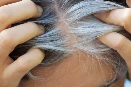
That persistent itchy patch might be eczema even if it doesn’t look like the textbook pictures you’ve seen online. For the millions of people with darker skin tones, atopic dermatitis often hides behind a disguise that many healthcare providers miss entirely. While medical literature has traditionally depicted eczema as red, inflamed patches, this description primarily applies to lighter skin. On darker complexions, the condition presents with subtle differences that frequently lead to misdiagnosis, delayed treatment, and unnecessary suffering.
Understanding how eczema uniquely appears on melanin-rich skin could be the key to finally addressing that mysterious skin issue your doctor keeps misidentifying. The distinctive signs might explain symptoms you’ve been experiencing for years without proper diagnosis or relief.
The invisible inflammation that doctors often miss
The classic description of eczema as “red, inflamed patches” creates a fundamental diagnostic challenge for individuals with darker skin. This standard characterization fails to acknowledge how inflammation manifests differently across the spectrum of skin tones.
In darker skin, inflammation doesn’t produce the telltale redness so prominently featured in medical textbooks and online resources. Instead, affected areas often appear as darker brown, ashen gray, or purplish patches that can be subtle and easily overlooked during brief medical examinations. This pigmentation difference represents the same inflammatory process occurring in lighter skin, but the visual cues differ dramatically due to how melanin interacts with inflammation.
The traditional inflammation markers that medical professionals learn to identify during training often focus on characteristics most visible in lighter skin. When these expected signs don’t appear as anticipated, even experienced providers sometimes miss clear cases of atopic dermatitis. This educational gap continues affecting diagnostic accuracy despite growing awareness of presentation differences across skin tones.
Textural changes often provide more reliable indicators than color changes in darker skin. The skin may feel rough, thickened, or bumpy when touched, even when visual cues appear minimal. This tactile difference highlights why thorough physical examination beyond visual inspection becomes crucial for accurate diagnosis in darker-skinned individuals.
Scratch marks and excoriations sometimes provide the first visible evidence that leads to proper diagnosis. These self-inflicted skin injuries result from the intense itching that characterizes eczema across all skin tones. When providers notice these scratch patterns but can’t identify obvious inflammation, it should trigger consideration of atopic dermatitis manifesting with skin-tone-specific characteristics.
The diagnostic confusion extends beyond just missing the condition entirely. Darker-skinned individuals with eczema frequently receive incorrect diagnoses for other conditions with similar appearances, including fungal infections, psoriasis, or contact dermatitis. These misdiagnoses lead to inappropriate treatments that fail to address the underlying condition while potentially causing additional skin irritation.
The telltale signs unique to melanin-rich skin
Several distinctive patterns and manifestations help identify atopic dermatitis in darker skin tones, providing crucial diagnostic clues when traditional signs prove absent or misleading.
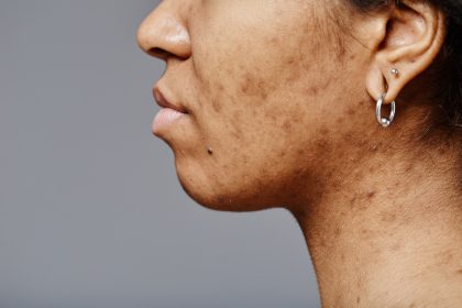
Hyperpigmentation, where affected skin becomes noticeably darker than surrounding areas, represents one of the most common eczema indicators in melanin-rich skin. This darkening occurs as inflammatory processes trigger melanocytes to increase pigment production, creating patches that may appear brown, dark purple, or almost black depending on the individual’s base skin tone. These hyperpigmented areas often persist long after the active inflammation subsides, creating lasting evidence of previous flares.
Conversely, hypopigmentation sometimes develops instead, particularly after chronic inflammation. These lighter patches result from inflammation damaging melanocytes or disrupting normal pigment production. The contrast between these lighter areas and surrounding unaffected skin creates distinctive patches that can persist for months or years even after successful eczema treatment, causing significant psychological impact beyond the physical symptoms.
Follicular prominence, where tiny bumps appear around hair follicles creating a persistent goosebump-like texture, occurs more commonly in darker skin. This presentation, medically termed “perifollicular accentuation,” creates a distinctive appearance that experienced dermatologists recognize as strongly associated with atopic dermatitis in melanin-rich skin. The bumps typically concentrate in characteristic eczema locations like the outer arms, thighs, and trunk.
Lichenification develops more readily and prominently in darker skin with chronic eczema. This thickening and exaggeration of normal skin lines creates a leathery, almost bark-like texture in areas subjected to persistent scratching and rubbing. The visual appearance resembles tree bark or leather, while feeling distinctly thickened and rough to the touch. This change results from the skin’s response to chronic irritation and commonly affects flexural areas like elbow and knee folds.
Prurigo nodularis, consisting of extremely itchy, hard, raised bumps, develops more frequently as a complication of atopic dermatitis in darker skin. These intensely pruritic nodules result from persistent scratching and can develop a cycle where itching leads to scratching, which creates more nodules and intensifies itching further. The distinctive firm, dome-shaped papules and nodules typically measure 3-20mm in diameter and often develop hyperpigmented borders with occasional scaling.
Papular eczema, where the rash appears primarily as small, distinct bumps rather than confluent patches, occurs more commonly in individuals of African and Caribbean descent. These widespread papules create a characteristic appearance that differs significantly from the diffuse patches typically depicted in eczema educational materials. Recognizing this presentation pattern proves essential for accurate diagnosis in affected populations.
The hidden burden of persistent itch
While visual differences in eczema presentation create diagnostic challenges, the subjective experience of intense, persistent itching remains consistent across all skin tones. This symptom often proves even more debilitating for darker-skinned individuals due to several compounding factors.
Itch intensity frequently reaches more severe levels in darker skin, according to multiple research studies measuring pruritus in different populations. This increased severity may result from differences in inflammatory signaling, neurosensory processing, or skin barrier function related to genetic factors that correlate with melanin production. The practical impact means darker-skinned individuals often experience more extreme discomfort despite sometimes showing less visible inflammation.
The itch-scratch cycle creates particularly problematic consequences in darker skin. Scratching triggers both immediate and delayed hyperpigmentation that persists long after the itch subsides, creating visible evidence of the condition that may affect confidence and social comfort. This physical evidence often remains visible for months or years, essentially tattooing a record of the inflammatory episode onto the skin surface through altered pigmentation.
Sleep disruption severity increases with itch intensity, creating disproportionate impact on darker-skinned individuals who report higher average itch levels. This sleep degradation affects daytime functioning, work performance, and overall quality of life, extending the condition’s impact far beyond skin symptoms alone. The resulting fatigue can further compromise immune function, potentially triggering additional flares in a self-perpetuating cycle.
Psychological burden increases through multiple mechanisms when diagnosis and treatment delays occur due to presentation differences. The extended duration of uncontrolled symptoms, frustration with ineffective treatments, and distress over visible skin changes all contribute to higher rates of anxiety and depression among darker-skinned individuals with atopic dermatitis. This psychological impact often receives inadequate attention during treatment planning.
The visible evidence of scratching creates particular social challenges that may not receive appropriate acknowledgment from healthcare providers. Hyperpigmented patches and lichenification in visible areas like the face, neck, and hands can trigger social anxiety, self-consciousness, and even workplace discrimination. These real-world impacts deserve consideration as part of comprehensive treatment planning rather than focusing exclusively on physical symptoms.
The pigmentation aftermath most doctors never mention
Even after successful treatment controls active inflammation, darker-skinned individuals often face lasting pigmentation changes that persist long after the underlying eczema resolves. This post-inflammatory pigment alteration creates an extended burden rarely addressed in treatment discussions.
Post-inflammatory hyperpigmentation develops when inflammation triggers increased melanin production that continues even after the inflammatory process subsides. These darkened areas may persist for months to years following successful eczema treatment, creating a visible reminder of previous flares. The duration and intensity vary significantly between individuals, with some experiencing nearly permanent changes while others see gradual fading over time.
Post-inflammatory hypopigmentation, where affected areas become lighter than surrounding skin, occurs when inflammation damages melanocytes or disrupts normal pigment production. These lightened patches frequently cause greater psychological distress than hyperpigmentation, particularly in individuals with deeper skin tones where the contrast appears more pronounced. Unlike hyperpigmentation which may eventually fade, hypopigmented areas often remain permanently altered.
The psychological impact of these pigmentary changes frequently exceeds the distress caused by the original eczema symptoms for many individuals. The visible evidence of previous inflammation affects clothing choices, social comfort, and self-perception even when the physical discomfort of active eczema has resolved. This extended burden rarely receives adequate acknowledgment during treatment discussions.
Treatment options for post-inflammatory pigmentation remain limited and often prove less effective than desired. While various lightening agents, chemical peels, and laser therapies show some efficacy for hyperpigmentation, results vary widely between individuals. Hypopigmentation proves particularly resistant to treatment, with few reliable options for restoring normal pigmentation to affected areas.
Sun protection becomes especially crucial for preventing worsening of pigmentary changes, though this important aspect of care often receives insufficient emphasis during treatment discussions. Ultraviolet exposure can darken hyperpigmented areas while potentially making hypopigmented regions more noticeable through tanning of surrounding skin. Consistent, appropriate sun protection helps minimize these effects while allowing gradual normalization of pigment over time.
The cumulative surface area affected by pigmentary changes typically increases over time with repeated eczema flares. Each inflammatory episode creates new regions of altered pigmentation that add to the existing pattern, gradually increasing the visible evidence of the condition. This accumulation underscores the importance of prompt, effective treatment that minimizes inflammation before pigmentary changes become established.
Finding solutions that actually work for your skin
Managing eczema effectively in darker skin requires approaches tailored to the specific challenges and manifestations that occur more commonly in melanin-rich skin. Several strategies prove particularly important for addressing these unique considerations.
Early intervention becomes especially crucial for preventing the pigmentary changes that disproportionately affect darker skin. Seeking treatment at the first signs of itching or subtle texture changes, rather than waiting for obvious visual symptoms, helps minimize inflammation before it triggers lasting pigmentation alterations. This proactive approach often requires self-advocacy when healthcare providers might otherwise dismiss early symptoms as insignificant based on limited visible changes.
Appropriate corticosteroid selection requires special consideration for darker skin. Medium to high-potency topical steroids sometimes prove necessary to adequately control inflammation despite subtle visual signs, particularly when significant itching suggests more intense underlying inflammation than visibly apparent. However, steroid-induced hypopigmentation poses greater cosmetic concern in darker skin, requiring careful monitoring and appropriate steroid-sparing strategies to minimize this risk.
Calcineurin inhibitors like tacrolimus and pimecrolimus offer particular advantages for facial and neck eczema in darker skin. These non-steroidal anti-inflammatory treatments effectively reduce eczema symptoms without causing the hypopigmentation that topical steroids may produce. This benefit proves especially valuable for visible areas where pigmentary changes would create significant cosmetic concern.
Barrier repair emphasis takes on heightened importance in darker skin, which research suggests may have inherently greater transepidermal water loss compared to lighter skin tones. Consistent use of appropriate moisturizers, particularly those containing ceramides, helps restore and maintain skin barrier function while reducing inflammation triggers. This fundamental approach often receives insufficient emphasis compared to anti-inflammatory treatments despite its crucial importance.
Trigger identification through systematic tracking helps identify specific factors that provoke flares in individual cases. Common triggers include certain fabrics, detergents, soaps, foods, stress, and environmental factors like low humidity or heat. Recognizing and avoiding personal triggers often provides more sustainable improvement than relying exclusively on medications to control symptoms after flares begin.
Post-inflammatory pigmentation management deserves specific attention as part of comprehensive treatment. Adding ingredients like niacinamide, vitamin C, azelaic acid, or licorice extract to skincare routines may help address hyperpigmentation, while sun protection prevents worsening of both hyper and hypopigmentation. These approaches work best when implemented early, ideally alongside anti-inflammatory treatment rather than waiting until pigment changes become established.
Building your healthcare team for better outcomes
Finding appropriate medical care for eczema in darker skin often requires strategic approaches to overcome systemic knowledge gaps and potential biases that affect diagnosis and treatment quality.
Dermatologist selection deserves careful consideration when seeking care for atopic dermatitis in darker skin. While any board-certified dermatologist should theoretically possess the knowledge to treat diverse skin tones, the reality often falls short of this ideal. Seeking providers with specific experience treating patients with similar skin tones, whether through direct questioning during appointment scheduling, reviewing provider photos on practice websites, or consulting community recommendations, often leads to more accurate diagnosis and appropriate treatment recommendations.
Preparation for appointments significantly impacts diagnostic accuracy and treatment quality. Bringing photos of affected areas during both flare and non-flare periods helps providers visualize the condition’s full presentation even when active inflammation appears minimal during the visit. Documenting itch severity, sleep disruption, and emotional impact provides crucial context beyond visible skin changes that might otherwise receive insufficient attention.
Clear communication about pigmentary concerns proves essential for comprehensive treatment planning. Explicitly discussing fears about lasting skin darkening or lightening ensures these issues receive appropriate consideration rather than focusing exclusively on controlling active inflammation. This conversation should include realistic expectations about potential pigmentary outcomes and available management options to address these changes if they occur.
Requesting skin-tone-appropriate education materials helps reinforce proper home care through visual resources that accurately represent how the condition appears in similar skin. While such materials remain unfortunately limited, some organizations and practices have developed resources specifically showing eczema manifestations across diverse skin tones. Having visual references that match your own presentation improves recognition of early flare signs and treatment response assessment.
Follow-up persistence ensures adequate monitoring of both inflammatory control and pigmentary changes. The extended timeline for pigmentation normalization after inflammation resolves necessitates longer-term follow-up than might be typical for lighter-skinned patients. Advocating for continued care until both active symptoms and pigmentary aftermath have adequately resolved helps prevent premature treatment discontinuation.
Second opinions warrant consideration when initial treatment provides inadequate relief or when providers seem unfamiliar with eczema’s unique presentation in darker skin. Different dermatologists bring varying levels of experience with diverse skin tones, and seeking alternative perspectives often leads to more effective treatment approaches for cases initially misdiagnosed or inadequately managed.
The landscape of dermatological care for darker skin continues evolving, with growing recognition of the need for more inclusive training, research, and clinical approaches. By combining informed self-advocacy with strategic healthcare team development, individuals with darker skin can overcome historical diagnostic and treatment disparities to achieve better eczema management and improved quality of life.