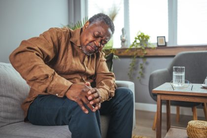
That distinctive pain or discomfort at the back of your knee can range from mildly annoying to severely debilitating. While often dismissed as a minor issue that will resolve on its own, posterior knee pain frequently signals underlying problems requiring proper attention. Understanding the various causes, recognizing important warning signs, and knowing when to seek medical care can help you address this common complaint effectively and prevent potential complications.
The anatomy behind the pain
The posterior knee region contains a complex arrangement of structures, any of which can become sources of pain when injured or inflamed. Understanding this anatomy helps explain the various ways discomfort can manifest in this area.
Muscles surrounding the back of the knee include the hamstrings (semitendinosus, semimembranosus, and biceps femoris) that run down the back of the thigh and connect behind the knee. These powerful muscles flex the knee and extend the hip, making them crucial for walking, running, and jumping activities. The gastrocnemius muscle, which forms the upper part of the calf, also originates above the knee joint and crosses behind it. Additionally, the small but important popliteus muscle sits specifically behind the knee joint and helps unlock the knee during bending movements.
Tendons connect these muscles to bones in the area, with the hamstring tendons attaching to the upper tibia and fibula (lower leg bones), while the gastrocnemius attaches to the lower femur (thigh bone). These tendons transmit force from muscles to bones but can become irritated or damaged with overuse or injury. The popliteus tendon plays a specialized role in knee rotation and stability.
Ligaments provide crucial stability to the knee joint, with the posterior cruciate ligament (PCL) being the primary ligament at the back of the knee. The PCL prevents excessive backward movement of the tibia relative to the femur and can be injured during direct impacts to the front of the knee. Other important ligaments include portions of the lateral and medial collateral ligaments that help stabilize the sides of the knee but have components that extend posteriorly.
The joint capsule surrounds the entire knee joint, with its posterior portion containing specialized pouches and potential spaces. One such space can fill with fluid and form a Baker’s cyst (popliteal cyst) behind the knee when joint irritation or injury occurs. The posterior capsule also contains nerve endings that can generate pain signals when stretched or inflamed.
Bursae are small fluid-filled sacs that reduce friction between moving structures. The popliteal bursa sits behind the knee and can become inflamed (bursitis), causing localized pain and swelling. Other bursae in the area include the semimembranosus bursa and the medial gastrocnemius bursa, which can similarly become irritated.
Blood vessels and nerves running through the posterior knee include the popliteal artery and vein, which are the main vessels supplying the lower leg, along with branches of the sciatic nerve. These structures pass through the popliteal fossa (the diamond-shaped space behind the knee) and can be sources of pain if compressed, damaged, or affected by conditions like blood clots.
Understanding this complex anatomy explains why many different conditions can cause similar pain patterns in the posterior knee, making accurate diagnosis sometimes challenging but essential for proper treatment.
Common causes of posterior knee pain
Posterior knee pain stems from various conditions affecting different structures, each with distinctive patterns of onset, symptoms, and appropriate treatments.
Muscle strains frequently cause pain behind the knee, particularly affecting the hamstrings or gastrocnemius muscles. These strains typically occur during sudden acceleration or deceleration movements, such as sprinting or abruptly stopping while running. The pain often develops suddenly with a characteristic “pulling” sensation and may be accompanied by muscle weakness, swelling, and localized tenderness. Mild strains (Grade 1) cause minimal tear of muscle fibers, while more severe strains (Grades 2 and 3) involve significant or complete muscle tearing. People with inadequate warm-up, previous muscle injuries, or muscle imbalances face higher risk of experiencing these strains.
Tendonitis or tendinopathy involves inflammation or degeneration of tendons behind the knee, commonly affecting the hamstring tendons, popliteus tendon, or gastrocnemius tendon. Unlike sudden strains, these conditions typically develop gradually from repetitive stress or overuse activities. Runners, cyclists, and individuals who frequently climb stairs often develop these conditions, experiencing pain that worsens with activity and improves with rest. The affected tendon may feel tender to touch, with noticeable stiffness after periods of inactivity, particularly in the morning or after sitting for extended periods.
Baker’s cysts (popliteal cysts) form when joint fluid accumulates in the back of the knee due to underlying inflammation or injury within the joint. These fluid-filled sacs create a noticeable bulge behind the knee that may become painful when fully flexing or extending the knee. People often describe a feeling of pressure or tightness rather than sharp pain. These cysts commonly occur alongside other knee conditions like arthritis or meniscus tears and may occasionally rupture, causing sudden calf pain and swelling that can mimic deep vein thrombosis.
Ligament injuries, particularly to the posterior cruciate ligament (PCL), cause significant posterior knee pain. PCL injuries typically result from direct impact to the front of the bent knee, such as hitting the dashboard in a car accident or falling directly onto the bent knee. Unlike the more commonly injured anterior cruciate ligament (ACL), PCL injuries often don’t produce an audible “pop” but create immediate pain and swelling behind the knee. These injuries frequently lead to knee instability, particularly when walking downstairs or on declining surfaces.
Meniscus tears affecting the posterior portions of these cushioning cartilages can cause pain localized to the back of the knee. These tears commonly occur during twisting movements while the knee bears weight, creating sudden pain often accompanied by clicking, locking, or catching sensations. The posterior horn of the medial meniscus is particularly vulnerable to tearing, especially in older adults with degenerative changes or in athletes performing frequent pivoting movements.
Arthritis affecting the knee joint can cause posterior pain, particularly when inflammation affects the back portion of the joint. Osteoarthritis, rheumatoid arthritis, and post-traumatic arthritis can all manifest with posterior knee pain, typically accompanied by stiffness, swelling, and pain that worsens with activity and improves with rest. Morning stiffness lasting more than 30 minutes particularly suggests inflammatory arthritis conditions. These arthritic conditions generally produce more diffuse knee pain rather than specifically posterior pain, but the posterior aspect may become the dominant pain location in some individuals.
Deep vein thrombosis (DVT) represents the most serious cause of posterior knee pain, occurring when blood clots form in deep veins, commonly in the calf or thigh. DVT often creates pain behind the knee where major blood vessels pass through the popliteal space. Unlike mechanical causes of knee pain, DVT pain typically occurs without injury and may be accompanied by swelling, warmth, and redness. Risk factors include prolonged immobility (such as long flights or bed rest), recent surgery, pregnancy, hormonal medications, and certain medical conditions. DVT requires immediate medical attention due to the risk of pulmonary embolism if clots break loose and travel to the lungs.
When to seek immediate medical attention
While many causes of posterior knee pain can be managed conservatively, certain symptoms require urgent medical evaluation due to potentially serious underlying conditions.
Sudden, severe pain behind the knee without injury, especially when accompanied by swelling, warmth, and redness, should trigger immediate medical consultation. This symptom pattern raises concerns about deep vein thrombosis (DVT), particularly if accompanied by risk factors such as recent surgery, prolonged immobility, pregnancy, or family history of clotting disorders. DVT represents a medical emergency due to the risk of pulmonary embolism, a life-threatening condition occurring when clots break loose and travel to the lungs.
Inability to bear weight on the affected leg warrants urgent evaluation, as this suggests significant structural damage. While minor strains or tendonitis may cause discomfort with walking, complete inability to support weight indicates more serious injury such as complete tendon rupture, severe ligament tear, or fracture affecting the knee joint. Delaying treatment for these conditions can lead to improper healing, prolonged recovery time, and potential long-term disability.
Visible deformity or significant swelling of the knee requires prompt medical assessment. Abnormal appearance suggests structural damage such as dislocation, fracture, or complete ligament/tendon rupture. Significant swelling, particularly when developing rapidly within hours of injury, indicates substantial internal bleeding or joint effusion requiring professional evaluation and management. Both conditions may need immobilization, drainage, or surgical intervention depending on severity.
Fever accompanied by knee pain and warmth raises concerns about infection within or around the knee joint (septic arthritis or cellulitis). Infections in this region can spread rapidly and cause permanent joint damage if not treated promptly with appropriate antibiotics and potentially surgical drainage. People with compromised immune systems, recent injuries breaking the skin, or previous joint replacements face particular risk for these infections.
Numbness or tingling extending below the knee suggests nerve involvement that requires immediate attention. These symptoms may indicate pressure on nerves from swelling, direct nerve injury, or conditions affecting the spine that manifest as posterior knee pain. Prolonged nerve compression can lead to permanent nerve damage if not addressed promptly, potentially resulting in chronic pain, weakness, or sensory changes.
Popping or snapping sounds at the time of injury followed by immediate pain and swelling typically indicate significant structural damage such as ligament tears or meniscus injuries. While not always requiring emergency care, these injuries should be evaluated within 24-48 hours to determine their severity and appropriate treatment plan. Delaying evaluation may complicate treatment and prolong recovery time.
When these warning signs appear, especially in combination, don’t attempt to self-diagnose or manage the condition at home. Prompt medical evaluation provides the best opportunity for accurate diagnosis, appropriate treatment, and optimal recovery from potentially serious conditions affecting the posterior knee.
Diagnosis: Identifying the source of pain
Healthcare providers use a systematic approach to identify the specific cause of posterior knee pain, employing various diagnostic tools and assessments to ensure accurate diagnosis and appropriate treatment planning.
The medical history assessment forms the foundation of diagnosis, with providers asking detailed questions about pain onset, duration, and characteristics. Specific queries typically include when the pain began, whether it started suddenly or gradually, activities that worsen or improve the pain, and any history of previous knee injuries or conditions. The provider will also inquire about occupation, sports participation, and other activities that might contribute to knee stress. This comprehensive history often provides crucial clues about the underlying cause before any physical examination begins.
Physical examination includes several structured assessments designed to evaluate different knee structures. The examination typically begins with observation of gait (walking pattern) and knee appearance, looking for visible swelling, bruising, or deformity. Range of motion testing evaluates how completely the knee can bend and straighten, noting any restrictions or pain during these movements. Specific tests for each structure include hamstring tension assessment, ligament stability tests like the posterior drawer test (for PCL integrity), and palpation of tendons and muscles to identify specific tender areas. The provider will also examine areas above and below the knee, as conditions affecting the hip or ankle can sometimes manifest as posterior knee pain.
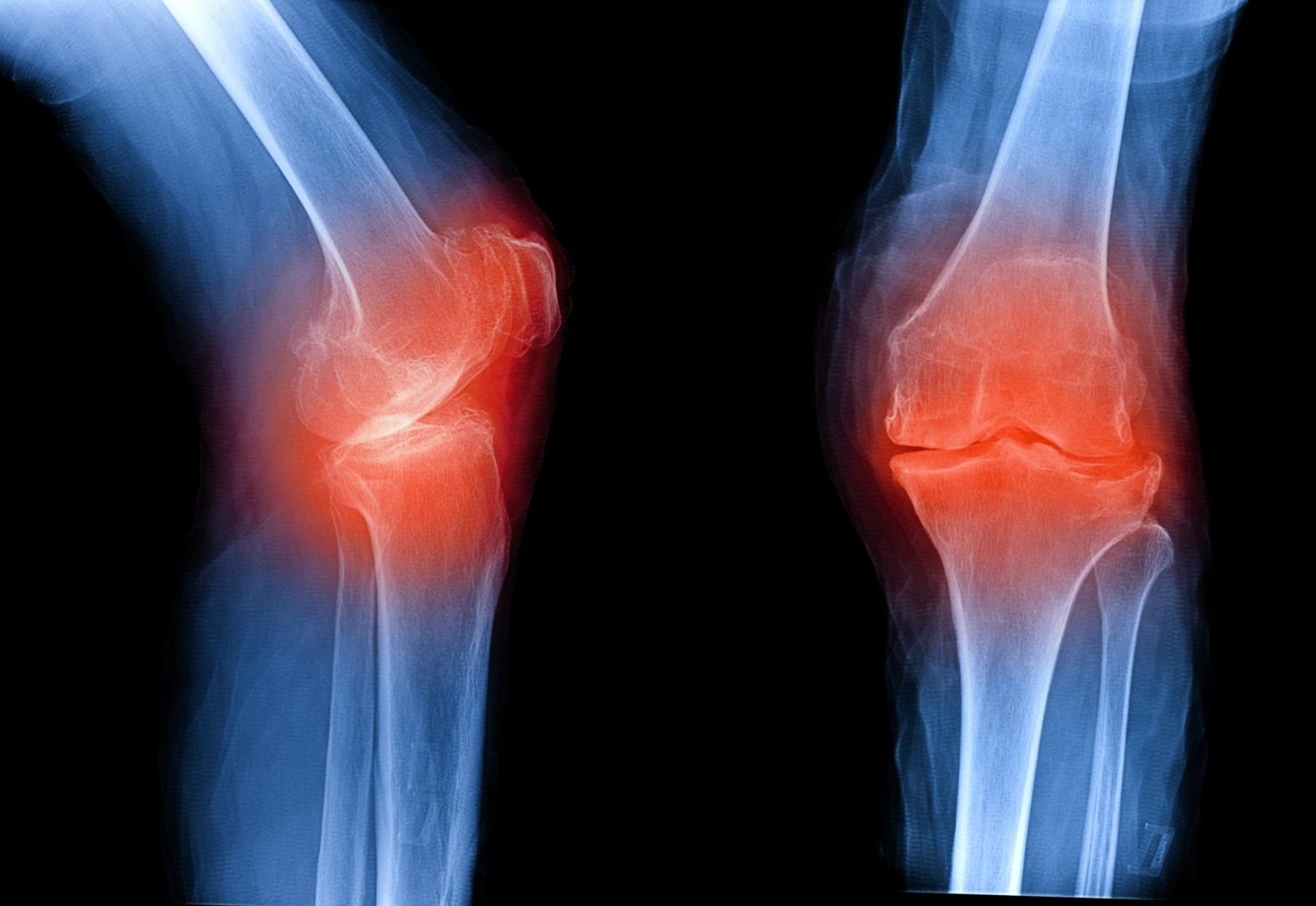
Imaging studies provide visualization of internal knee structures when the diagnosis remains unclear after history and physical examination. X-rays offer excellent visualization of bones, helping identify fractures, arthritis, or bone spurs, though they don’t show soft tissues like muscles, tendons, and ligaments. Magnetic Resonance Imaging (MRI) provides detailed images of all knee structures and represents the gold standard for diagnosing soft tissue injuries like meniscus tears, ligament damage, and tendon injuries. Ultrasound offers real-time visualization of tendons, muscles, and cysts, with the advantage of allowing dynamic assessment during movement, though with less detail than MRI. For suspected blood vessel problems like DVT, Doppler ultrasound specifically evaluates blood flow through vessels behind the knee.
Additional diagnostic procedures may be necessary in complex cases. Aspiration (removing fluid from the knee joint with a needle) allows analysis of joint fluid for infection, inflammation, or blood when these conditions are suspected. Electromyography (EMG) and nerve conduction studies evaluate nerve function when nerve-related symptoms like numbness or tingling accompany posterior knee pain. In cases where DVT is suspected, D-dimer blood tests may be ordered, though these are often followed by imaging studies for confirmation since D-dimer levels can be elevated for various reasons.
The diagnostic process often proceeds iteratively, with initial assessments guiding the selection of additional tests as needed. This systematic approach ensures accurate identification of the underlying cause, which is essential for developing an effective treatment plan tailored to the specific condition affecting the posterior knee.
Treatment approaches for posterior knee pain
Treatment strategies for posterior knee pain vary significantly depending on the underlying cause, severity of symptoms, and individual patient factors. Understanding these approaches helps patients participate actively in their recovery process.
Conservative management represents the first-line treatment for most cases of posterior knee pain, particularly those resulting from minor injuries or overuse conditions. The RICE protocol (Rest, Ice, Compression, Elevation) forms the foundation of initial treatment for acute injuries. This approach reduces inflammation and pain during the early healing phase, typically the first 48-72 hours after injury. Activity modification involves temporarily avoiding movements that aggravate symptoms while maintaining appropriate activity levels to prevent deconditioning. Over-the-counter pain medications like nonsteroidal anti-inflammatory drugs (NSAIDs) help manage pain and reduce inflammation, though they should be used as directed and with awareness of potential side effects, particularly with prolonged use.
Physical therapy plays a crucial role in recovery from most posterior knee conditions. Therapeutic exercises focus on gradually restoring strength to muscles supporting the knee, particularly the hamstrings, quadriceps, and calf muscles. Flexibility training improves range of motion and reduces tension in tight muscles that may contribute to pain. Manual therapy techniques performed by physical therapists, including soft tissue mobilization, joint mobilizations, and specialized massage, help decrease pain and improve tissue mobility. Modalities such as ultrasound, electrical stimulation, or iontophoresis may complement exercise-based approaches by reducing pain and promoting healing. Physical therapists also provide education about proper mechanics during daily activities and sports to prevent recurrence of symptoms.
Bracing and supportive devices assist recovery for certain conditions. Compression sleeves provide mild support while reducing swelling in many posterior knee conditions. More structured bracing may be recommended for ligament injuries, with specific braces designed to limit harmful movements while allowing functional activities. Kinesiology taping techniques can provide proprioceptive feedback and mild support for muscles and tendons while allowing full range of motion. Orthotic inserts sometimes help address biomechanical issues contributing to posterior knee pain, particularly when abnormal foot mechanics place additional stress on knee structures.
Advanced interventional treatments may be recommended when conservative approaches provide insufficient relief. Corticosteroid injections delivered precisely to inflamed tissues can provide significant temporary pain relief and inflammation reduction, though their use is typically limited due to potential side effects with repeated injections. Platelet-rich plasma (PRP) injections, derived from the patient’s own blood, deliver concentrated growth factors to injured tissues to promote healing, particularly for chronic tendon injuries. Hyaluronic acid injections may help lubricate the joint and reduce pain in cases where arthritis contributes to posterior knee symptoms. These interventional approaches are typically performed under ultrasound or fluoroscopic guidance to ensure accurate placement.
Surgical intervention becomes necessary in cases with significant structural damage or when conservative treatments fail to provide adequate relief. Arthroscopic surgery uses small incisions and specialized instruments to address issues like meniscus tears or loose bodies in the joint while minimizing tissue damage and recovery time. Ligament reconstruction may be required for complete tears of the posterior cruciate ligament, particularly in younger, active individuals. Tendon repair procedures address complete or severe partial tears of tendons like the hamstring or gastrocnemius. More extensive open surgical procedures might be necessary for complex injuries involving multiple structures or for joint replacement in cases of severe arthritis.
Treatment for medical conditions causing posterior knee pain, such as deep vein thrombosis, focuses on the underlying condition rather than knee symptoms specifically. DVT typically requires anticoagulant medications to prevent clot growth and reduce the risk of pulmonary embolism, sometimes beginning with injectable medications followed by oral anticoagulants. For septic arthritis (joint infection), intravenous antibiotics and surgical drainage of the joint are typically required to prevent permanent joint damage.
The optimal treatment approach often involves combining multiple strategies tailored to the specific diagnosis, symptom severity, and individual patient factors like age, activity level, and overall health. Close collaboration between patients and healthcare providers ensures that treatment plans address both immediate symptom relief and long-term functional restoration.
Prevention strategies for posterior knee pain
While not all causes of posterior knee pain can be prevented, several evidence-based strategies can significantly reduce the risk of developing these conditions, particularly for active individuals and those with previous knee issues.
Proper warm-up before physical activity prepares muscles, tendons, and joints for the stresses of exercise, reducing injury risk. An effective warm-up should include 5-10 minutes of light cardiovascular activity to increase blood flow to muscles, followed by dynamic stretching movements that mimic the planned activity. Static stretching (holding stretches in a fixed position) is more appropriate after exercise when tissues are already warm. This preparation process increases tissue flexibility and joint lubrication while enhancing neuromuscular coordination, all factors that help prevent injuries to posterior knee structures.
Strengthening exercises for muscles supporting the knee create a balanced foundation that reduces injury risk. Hamstring strengthening deserves particular attention for posterior knee health, with exercises like hamstring curls, bridges, and Romanian deadlifts developing these muscles effectively. Quadriceps strengthening provides balanced support from the front, while calf strengthening supports structures below the knee. Core strengthening improves overall body mechanics during activity, reducing inappropriate stresses on the knee joint. These exercises should progress gradually in intensity and be performed with proper form to maximize benefits while minimizing injury risk.
Flexibility maintenance through regular stretching helps prevent the muscle tightness that often contributes to posterior knee problems. Hamstring stretching receives particular emphasis, with techniques like seated forward bends or standing stretches performed regularly to maintain optimal muscle length. Calf stretching, including both gastrocnemius and soleus components, prevents tightness that can alter knee mechanics. Quadriceps stretching completes a balanced approach to knee flexibility. Holding stretches for 30 seconds and performing them when muscles are warm maximizes effectiveness while minimizing injury risk.
Proper technique during activities reduces abnormal stresses on knee structures that can lead to pain and injury. Maintaining proper alignment during squatting movements keeps the knees tracking over the toes rather than collapsing inward, which places excessive stress on ligaments and tendons. Landing mechanics when jumping should emphasize soft landings with bent knees to absorb impact forces gradually rather than suddenly. Running technique should include appropriate stride length and foot strike patterns that minimize impact forces transferred to the knees. For workplace activities, proper lifting techniques and ergonomic considerations help prevent occupational stresses to knee structures.
Appropriate footwear provides the foundation for proper lower extremity mechanics and shock absorption. Activity-specific shoes designed for individual sports or activities offer appropriate support and cushioning for those movements. Regular replacement of athletic shoes before they lose their supportive properties prevents the biomechanical changes that can contribute to knee problems. For individuals with specific foot mechanics like overpronation or high arches, shoes with appropriate support features or custom orthotics may provide additional protection against knee issues by optimizing overall lower limb alignment.
Training modifications help prevent overuse injuries that commonly affect posterior knee structures. The “10% rule” suggests limiting increases in training volume or intensity to no more than 10% weekly, allowing tissues to adapt gradually to increasing demands. Cross-training incorporates various activities that stress different body systems and structures, reducing repetitive strain on specific knee components. Periodization approaches systematically vary training intensity and volume throughout the year, incorporating planned recovery periods that allow tissues to heal and strengthen. For runners specifically, incorporating rest days, varying running surfaces, and gradually introducing hill or speed work helps prevent posterior knee issues common in this population.
Maintaining healthy body weight reduces stress on all knee structures, with research showing that each additional pound of body weight increases forces on the knee joint by 2-4 pounds during weight-bearing activities. Weight management through balanced nutrition and regular physical activity helps prevent the onset or progression of conditions like osteoarthritis that can manifest as posterior knee pain.
These prevention strategies provide a comprehensive approach to maintaining posterior knee health. While they require consistent effort and attention, they represent a worthwhile investment in avoiding the pain, activity limitations, and potential treatments required when posterior knee problems develop.
Living with posterior knee pain
For individuals with persistent or recurrent posterior knee pain, developing strategies for daily management helps maintain function and quality of life while working toward longer-term improvement.
Activity modification represents a crucial aspect of daily management, focusing on maintaining function while avoiding symptom exacerbation. This approach involves identifying specific movements or positions that consistently worsen symptoms and finding alternative ways to accomplish necessary tasks. For example, avoiding deep squatting or kneeling might be necessary for someone with a Baker’s cyst, while limiting stair climbing might help someone with patellar tendonitis. Importantly, activity modification differs from complete rest, as maintaining appropriate activity levels prevents the deconditioning that often worsens knee problems. Working with healthcare providers to develop individualized guidelines helps balance activity restrictions with necessary movement.
Pain management techniques help individuals function during daily activities while healing progresses. Ice application for 15-20 minutes several times daily helps reduce inflammation and pain, particularly after activities that stress the knee. Heat therapy, often most effective in chronic conditions, improves blood flow and relaxes tight muscles contributing to posterior knee discomfort. Over-the-counter medications like acetaminophen or NSAIDs provide temporary relief, though they should be used according to package directions and with awareness of potential side effects with prolonged use. Topical analgesics, including creams, gels, or patches containing ingredients like menthol, camphor, or NSAIDs, offer localized relief with fewer systemic effects than oral medications.
Supportive devices provide stability and reduce stress on painful structures during daily activities. Compression sleeves offer mild support while helping control swelling, making them suitable for many conditions affecting the posterior knee. More structured braces might be recommended for specific conditions, particularly those involving ligament injuries or instability. Kinesiology tape applied in specific patterns can provide proprioceptive feedback and gentle support while allowing full movement. For some individuals, walking aids like canes or trekking poles temporarily redistribute weight away from the affected leg during particularly symptomatic periods.
Ergonomic adjustments in home and work environments can significantly reduce stress on knee structures. Workplace modifications might include adjusting chair height to ensure proper knee positioning, using anti-fatigue mats for standing tasks, or reorganizing workspaces to minimize knee-stressing movements like deep squatting or kneeling. Home adjustments could include installing grab bars in bathrooms, using reaching tools to avoid bending, or rearranging frequently used items to avoid excessive knee stress during daily activities. These environmental changes, while sometimes simple, can substantially reduce cumulative stress on sensitive knee structures.
Self-management education empowers individuals to actively participate in their recovery and ongoing knee health. Understanding the specific condition causing posterior knee pain helps individuals recognize activities that might aggravate symptoms and identify early warning signs of potential flare-ups. Learning proper body mechanics for daily activities like lifting, carrying, and household tasks prevents additional stress on knee structures. Developing strategies for activity pacing helps maintain function while avoiding symptom exacerbation. This knowledge-based approach converts patients from passive recipients of care to active participants in their own health management.
Maintaining overall physical conditioning while working around knee limitations prevents the deconditioning that often worsens knee problems. Low-impact cardiovascular activities like swimming, water aerobics, or stationary cycling maintain cardiovascular fitness without excessive knee stress. Upper body and core strengthening exercises preserve overall strength and body mechanics while allowing knee healing. Developing modified exercise routines with guidance from healthcare providers ensures continued physical activity that supports rather than hinders recovery.
Psychological approaches address the emotional and cognitive aspects of living with posterior knee pain. Stress management techniques like deep breathing, meditation, or progressive muscle relaxation help reduce muscle tension that can exacerbate knee symptoms. Cognitive-behavioral strategies help individuals develop realistic expectations, focus on progress rather than limitations, and maintain motivation during recovery. For those with chronic pain, working with mental health professionals specializing in pain management can provide additional tools for coping with persistent symptoms while maintaining quality of life.
By integrating these various approaches to daily management, individuals with posterior knee pain can maintain function and quality of life while working toward long-term improvement through appropriate medical care and rehabilitation.
The road to recovery
Understanding the typical recovery process for posterior knee pain helps patients develop realistic expectations and recognize appropriate progress benchmarks as healing occurs.
Recovery timelines vary significantly depending on the specific condition causing posterior knee pain. Mild muscle strains typically improve within 2-4 weeks with appropriate rest and rehabilitation. Tendonitis conditions often require 6-12 weeks for substantial improvement, with longer recovery periods for tendinosis (degenerative tendon changes). Ligament injuries like PCL tears may take 3-9 months for recovery, depending on severity and whether surgical intervention was required. Meniscus injuries typically require 4-8 weeks for recovery from small tears treated conservatively, while surgical repair may require 3-6 months for full return to activities. Medical conditions like deep vein thrombosis may resolve with anticoagulant treatment within weeks, but ongoing management may be necessary to prevent recurrence.
Healing phases follow a relatively predictable pattern regardless of the specific condition. The acute phase typically lasts 1-7 days and involves controlling inflammation, protecting injured structures, and minimizing additional damage. The subacute phase follows for approximately 1-6 weeks (depending on the condition) and focuses on gradually restoring range of motion and beginning gentle strengthening as pain allows. The remodeling phase begins around 3-6 weeks post-injury and continues for months, focusing on progressive strengthening, endurance training, and gradual return to normal activities. Understanding these phases helps patients recognize that certain symptoms and limitations are normal parts of the recovery process rather than signs of failed treatment.
Rehabilitation progression should follow evidence-based protocols tailored to the specific condition. Early stages typically emphasize pain control, gentle range of motion exercises, and isometric strengthening (muscle contractions without joint movement). As healing progresses, exercises advance to include concentric strengthening (muscle shortening under tension), eccentric training (controlled muscle lengthening), and eventually functional movements that mimic daily activities and sports requirements. This gradual progression allows tissues to adapt to increasing demands without reinjury. Patients should understand that attempting to advance too quickly through these stages often leads to setbacks rather than faster recovery.
Return to activity guidelines help individuals safely resume sports, work, and recreational activities without reinjury. These guidelines typically involve meeting specific criteria rather than simply waiting a predetermined time period. Common benchmarks include achieving full, pain-free range of motion, restoring at least 90% of normal strength compared to the unaffected leg, demonstrating proper movement patterns during functional tests, and progressing through sport-specific or work-specific training without symptom exacerbation. These objective measures ensure that tissues have healed sufficiently to withstand the demands of returning activities.
Potential complications that may arise during recovery include persistent pain, recurrent swelling, movement compensation patterns that stress other structures, and psychological factors like fear of reinjury that limit full recovery. Recognizing these potential complications allows for early intervention when progress plateaus unexpectedly. In some cases, reassessment by healthcare providers may be necessary to identify additional treatments or modifications to the rehabilitation approach.
Long-term management strategies after recovery help prevent recurrence of posterior knee pain. These strategies typically include maintenance exercises that continue to strengthen key muscle groups, activity modifications that minimize stress on vulnerable structures, and regular self-monitoring for early signs of potential problems. For some conditions, particularly those involving degenerative changes, ongoing management may be necessary even after symptoms resolve to prevent or delay symptom return.
The recovery process for posterior knee pain rarely follows a perfectly linear path. Most individuals experience periods of improvement followed by temporary plateaus or even minor setbacks. Understanding this normal pattern helps maintain realistic expectations and prevents discouragement during the recovery journey. With appropriate treatment, rehabilitation, and patience, most individuals with posterior knee pain can return to their desired activities with good function and minimal or no ongoing symptoms.
Posterior knee pain, while common and sometimes persistent, can typically be effectively addressed through proper diagnosis and treatment. Understanding the complex anatomy of this region, recognizing common causes and warning signs, seeking appropriate medical care, and actively participating in treatment and prevention strategies provides the best foundation for recovering from current problems and preventing future issues.
If you’re experiencing posterior knee pain, remember that early evaluation often leads to simpler treatment and faster recovery. Don’t hesitate to consult healthcare providers when symptoms persist beyond a few days of home management or when warning signs suggest potentially serious conditions. With proper care and attention to knee health, most individuals can maintain active, pain-free lifestyles throughout their lives.