Understanding the various pathways to partial paralysis helps with both prevention and early intervention
The fundamental disruption behind movement loss
Partial paralysis occurs when nerve signals between the brain and specific muscle groups become interrupted, resulting in weakness or inability to move certain body parts while others remain functional. This condition differs from total paralysis, where complete loss of movement and sensation affects entire regions of the body. The nervous system pathways involved in partial paralysis function like complex electrical circuits, with damage at any point potentially disrupting the transmission process essential for voluntary movement.
The nervous system consists of two primary components that can experience damage leading to partial paralysis. The central nervous system includes the brain and spinal cord, serving as the command center and main transmission highway. The peripheral nervous system encompasses all nerves branching throughout the body, delivering messages between muscles and the central system. Damage to either component can result in partial paralysis, though the recovery outlook and treatment approaches differ significantly between them.
Partial paralysis manifests through various patterns depending on the location and extent of nervous system damage. Monoplegia affects a single limb, while hemiplegia impacts one side of the body. Paraplegia involves both legs, and quadriplegia affects all four limbs to varying degrees. These patterns provide important diagnostic clues about the underlying cause and location of nervous system injury, helping guide treatment approaches and rehabilitation strategies.
The timeline of symptom development provides crucial information about potential causes. Sudden onset partial paralysis typically results from acute events like stroke or trauma, while gradual progression over weeks or months suggests degenerative conditions, tumors, or inflammatory processes. Fluctuating symptoms that come and go might indicate multiple sclerosis, pressure from herniated discs, or certain metabolic disorders that temporarily affect nerve function.
Vascular events that interrupt nerve communication
Stroke represents the leading cause of partial paralysis in adults, with approximately 800,000 Americans experiencing strokes annually. These vascular events occur when blood flow to brain tissue becomes blocked by a clot (ischemic stroke) or when a blood vessel ruptures (hemorrhagic stroke). The specific body regions affected depend entirely on which brain areas lose oxygen supply during the event. Most commonly, stroke causes hemiplegia—paralysis affecting one side of the body opposite to the brain hemisphere where the stroke occurred.
The time between stroke onset and medical intervention dramatically impacts recovery potential. The critical four-hour window for administering clot-dissolving medications represents the period when oxygen-deprived brain cells remain salvageable before permanent damage occurs. This narrow timeframe highlights the importance of recognizing early stroke warning signs, including facial drooping, arm weakness, and speech difficulties. Prompt treatment often prevents partial paralysis from becoming permanent.
Transient ischemic attacks (TIAs), sometimes called “mini-strokes,” temporarily interrupt blood flow to brain regions without causing permanent damage. These events serve as serious warning signs, with approximately one-third of TIA patients later experiencing a full stroke. The temporary paralysis associated with TIAs typically resolves within 24 hours but requires immediate medical attention to prevent more severe episodes. Risk reduction measures following TIAs can significantly decrease the likelihood of developing permanent partial paralysis.
Less commonly, vascular malformations can lead to partial paralysis through pressure effects or bleeding. Arteriovenous malformations (AVMs) represent abnormal tangles of blood vessels that can rupture, causing localized brain hemorrhage. Cavernous angiomas, collections of dilated blood vessels with thin walls, may also leak blood slowly, creating gradually increasing pressure on surrounding neural tissue. These conditions often cause intermittent or slowly progressing weakness before more severe paralysis develops.
Trauma-induced nervous system damage
Spinal cord injuries account for approximately 17,500 new cases of paralysis annually in the United States. These injuries most commonly result from motor vehicle accidents, falls, sports injuries, and acts of violence. The spinal cord contains vital nerve pathways connecting the brain to the body, with damage at different vertebral levels producing predictable patterns of paralysis. Injuries to the cervical spine (neck region) frequently cause quadriplegia affecting all four limbs, while thoracic or lumbar injuries typically result in paraplegia affecting the legs.
The severity of spinal cord injuries exists on a spectrum rather than as complete or incomplete categories. Partial spinal cord damage often leaves some pathways intact, allowing for varying degrees of preserved function and recovery potential. This variability explains why some individuals with similar-looking injuries on imaging studies may experience dramatically different functional outcomes. The pattern of preserved sensation often provides clues about which nerve pathways remain intact and which rehabilitation approaches might prove most beneficial.
Traumatic brain injuries (TBIs) cause partial paralysis through direct damage to movement control centers in the brain. Approximately 1.5 million Americans sustain TBIs annually, with causes ranging from falls and vehicle accidents to sports injuries and combat-related blast exposure. The frontal and parietal lobes contain crucial motor control regions, with damage to these areas often resulting in contralateral weakness or paralysis (affecting the opposite side of the body). Recovery from TBI-related paralysis depends on the injury severity, prompt treatment, and the brain’s neuroplasticity—its ability to form new neural connections.
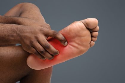
Peripheral nerve injuries often result from direct trauma such as lacerations, crushing injuries, or excessive stretching. These injuries frequently affect specific movement patterns corresponding to the damaged nerve’s territory. For example, radial nerve injury typically causes wrist drop, while peroneal nerve damage leads to foot drop. Many peripheral nerve injuries allow for significant recovery as these nerves can regenerate under the right conditions, though the process typically occurs slowly at a rate of approximately one inch per month.
Inflammatory and autoimmune attackers
Multiple sclerosis (MS) represents the most common inflammatory cause of partial paralysis in young adults. This condition occurs when the immune system mistakenly attacks myelin, the protective covering surrounding nerve fibers. These attacks create scarring (sclerosis) that disrupts nerve signal transmission, leading to weakness, numbness, and coordination problems that can progress to partial paralysis. The relapsing-remitting pattern characteristic of many MS cases creates periods of symptom improvement and worsening, with paralysis often fluctuating over time.
Guillain-Barré syndrome (GBS) causes rapidly progressing partial paralysis through immune-mediated attack on peripheral nerves. This rare condition typically begins with weakness in the legs that ascends to affect the arms and potentially breathing muscles. The disease often follows viral or bacterial infections, with the immune response triggering nerve damage. Most patients experience maximum weakness within two to four weeks, followed by a gradual recovery period lasting months. Early treatment with immunoglobulin therapy or plasma exchange significantly improves outcomes.
Transverse myelitis involves inflammation across a segment of the spinal cord, resulting in varying degrees of paralysis below the affected level. This condition can develop rapidly, often reaching maximum severity within hours to days. Approximately one-third of cases follow viral infections, while others associate with autoimmune disorders or occur without identifiable triggers. The inflammation damages nerve fibers crossing through the affected spinal segment, causing bilateral symptoms that may affect movement, sensation, and bowel/bladder function.
Chronic inflammatory demyelinating polyneuropathy (CIDP) represents a rare autoimmune condition affecting peripheral nerves throughout the body. Unlike Guillain-Barré syndrome’s acute presentation, CIDP develops gradually over at least eight weeks and often continues progressing for months or years without treatment. The condition typically causes symmetric weakness beginning in the legs and ascending to the arms, with periods of plateau or slight improvement followed by further deterioration. Early recognition and treatment with immunosuppressive therapies significantly improve the prognosis for maintaining mobility.
Space-occupying lesions that compress vital pathways
Brain tumors cause partial paralysis through pressure effects on motor control regions or their connecting pathways. Approximately 80,000 primary brain tumors are diagnosed annually in the United States, with metastatic tumors from other cancer sites occurring even more frequently. The gradual growth of these space-occupying lesions typically produces slowly progressing weakness before more severe paralysis develops. The pattern of motor deficit provides clues about tumor location, with modern neuroimaging techniques allowing precise localization before treatment.
Spinal tumors affect approximately 10,000 Americans annually and frequently cause partial paralysis through compression of spinal cord pathways. These tumors may originate in the spinal cord itself (intramedullary), in the protective membranes surrounding it (intradural extramedullary), or in the surrounding vertebrae (extradural). The growth rate significantly influences symptom progression, with rapidly growing tumors causing more abrupt paralysis. Night pain that worsens when lying down represents a characteristic symptom of many spinal tumors, often preceding weakness or paralysis by months.
Herniated discs represent common causes of nerve compression leading to focal weakness or partial paralysis. These injuries occur when the cushioning discs between vertebrae rupture, allowing the inner gel-like material to protrude and press against nearby nerve roots. Cervical disc herniations typically cause arm weakness corresponding to the affected nerve level, while lumbar herniations more commonly affect leg strength. The sudden nature of many disc herniations explains why patients often report weakness developing shortly after feeling a “pop” or immediate pain during lifting or twisting movements.
Abscesses developing within the spinal canal create urgent compression situations requiring immediate treatment. These infections typically spread from nearby structures like vertebrae (in cases of osteomyelitis) or from distant infection sites through bloodstream seeding. The abscess formation creates both direct pressure effects and inflammatory damage to neural structures. Unlike tumors, spinal abscesses often cause relatively rapid symptom progression over days rather than weeks or months, frequently accompanied by fever, severe pain, and elevated inflammatory markers in blood tests.
Developmental and congenital conditions
Cerebral palsy represents the most common developmental cause of partial paralysis, affecting approximately 1 in 345 children in the United States. This condition results from abnormal brain development or damage occurring before birth, during delivery, or in early infancy. The resulting movement disorders manifest as spastic paralysis (stiff, difficult movement), dyskinetic symptoms (uncontrolled movements), ataxic features (poor coordination), or mixed presentations. Though non-progressive, the movement limitations may appear to change during growth periods as affected muscles develop differently from their counterparts.
Spina bifida occurs when the spinal column fails to close completely during embryonic development, leaving portions of the spinal cord vulnerable to damage. This condition affects approximately 1,500 newborns annually in the United States, with severity ranging from asymptomatic hidden forms to severe cases with significant paralysis. The level of the spinal defect determines which body regions experience paralysis, with higher lesions causing more extensive movement limitations. Modern prenatal surgical intervention has improved outcomes for many affected children.
Chiari malformations involve structural abnormalities where brain tissue extends into the spinal canal, potentially compressing the spinal cord and causing progressive weakness or paralysis. Type I malformations, the most common form, often remain asymptomatic until adolescence or adulthood when symptoms emerge gradually. Headaches worsening with coughing or straining typically precede neurological symptoms like partial paralysis, numbness, or coordination problems. Surgical decompression often prevents symptom progression when performed before permanent nerve damage occurs.
Tethered cord syndrome develops when the spinal cord abnormally attaches to surrounding tissues, limiting its normal movement within the spinal canal. As children grow, this tethering creates progressive stretching and tension on the spinal cord, leading to neurological deterioration including partial paralysis. Early signs often include back pain, leg weakness, gait abnormalities, and bowel or bladder dysfunction. Surgical release of the tethered section prevents further deterioration but may not reverse existing paralysis, highlighting the importance of early diagnosis.
Metabolic and toxic pathways to nerve damage
Diabetes represents a leading cause of nerve damage (neuropathy) that can progress to partial paralysis, particularly in limbs. Approximately 60-70% of diabetic patients develop some degree of nerve damage over time, with risk increasing with disease duration and poor glucose control. The condition typically begins with sensory symptoms like numbness or tingling before progressing to motor weakness, most commonly affecting the feet and legs in a “stocking” distribution. Diabetic mononeuropathy can also cause sudden paralysis of isolated nerves, including those controlling eye movements or facial expressions.
Heavy metal toxicity from lead, mercury, arsenic, and thallium can cause peripheral nerve damage leading to partial paralysis. Occupational exposure represents the most common source, though environmental contamination and some traditional medicines also create risk. These toxins disrupt nerve cell metabolism and damage myelin, the protective covering around nerve fibers. The resulting weakness typically develops symmetrically, beginning in the hands and feet before progressing proximally. Chelation therapy to remove the metals from the body can halt progression but may not reverse existing paralysis.
Nutritional deficiencies, particularly of B vitamins, create vulnerability to nerve damage and partial paralysis. Vitamin B12 deficiency, common in strict vegetarians, those with pernicious anemia, or after certain gastric surgeries, damages the spinal cord’s long pathways controlling movement and sensation. Thiamine (B1) deficiency, often associated with alcoholism or prolonged malnutrition, causes peripheral nerve damage that can progress to partial paralysis if left untreated. These deficiency states respond dramatically to appropriate supplementation when identified before permanent nerve damage occurs.
Certain medications carry risks for nerve damage potentially leading to partial paralysis. Chemotherapy drugs, particularly vincristine and paclitaxel, commonly cause dose-dependent peripheral neuropathy affecting movement and sensation. Some antibiotics, including fluoroquinolones like ciprofloxacin, carry small risks of peripheral nerve damage. Antiretroviral medications used for HIV treatment can trigger neuropathy in susceptible individuals. These medication-induced forms of paralysis often improve with dose adjustment or medication changes, though recovery may take months after the offending drug discontinuation.
Recognizing warning signs requiring urgent attention
Sudden weakness or inability to move a body part represents an emergency requiring immediate medical evaluation. This symptom, particularly when accompanied by slurred speech, facial drooping, or confusion, suggests possible stroke—a time-sensitive emergency where treatment delays significantly worsen outcomes. The FAST acronym (Face drooping, Arm weakness, Speech difficulties, Time to call emergency services) provides an important memory tool for recognizing stroke symptoms requiring urgent intervention.
Progressive weakness developing over days to weeks warrants prompt medical attention to identify potentially reversible causes of paralysis. This timeframe suggests conditions like Guillain-Barré syndrome, spinal cord compression, or inflammatory processes that respond best to early treatment. The pattern of weakness progression provides important diagnostic clues, with ascending patterns (beginning in the feet and moving upward) suggesting different causes than descending or asymmetric patterns.
Weakness accompanied by sensory changes like numbness, tingling, or pain helps narrow potential causes and determines urgency. The distribution of these sensory changes provides valuable diagnostic information, with dermatomal patterns (following specific nerve root territories) suggesting different causes than glove-and-stocking distributions or scattered sensory patches. Complete loss of sensation in affected areas generally indicates more severe nerve damage than partial sensory changes.
Other accompanying symptoms provide crucial context for evaluating paralysis causes. Bowel or bladder dysfunction alongside weakness suggests spinal cord involvement requiring urgent evaluation. Visual changes accompanying paralysis might indicate multiple sclerosis or certain brain lesions. Cognitive changes or speech difficulties point toward brain involvement rather than spinal or peripheral nerve causes. These associated symptoms help direct appropriate diagnostic testing and treatment approaches.
Prevention strategies for modifiable risk factors
Vascular risk factor management significantly reduces stroke likelihood, the leading cause of acquired partial paralysis. Controlling high blood pressure, the most influential modifiable stroke risk factor, reduces risk by approximately 30% for each 10mmHg systolic pressure reduction. Smoking cessation cuts stroke risk in half after five years, while managing diabetes, maintaining healthy cholesterol levels, and treating atrial fibrillation further reduces vulnerability. These preventive measures provide the most effective protection against paralysis for most adults.
Injury prevention strategies dramatically reduce traumatic causes of partial paralysis. Vehicle safety practices including seatbelt use, avoiding distracted or impaired driving, and following speed limits prevent many spinal cord and brain injuries. Proper sporting equipment, including helmets for cycling and contact sports, reduces concussion risk. Fall prevention measures, particularly important for older adults, include home modification, balance training, and medication review to eliminate unnecessary drugs that increase fall risk.
Nerve protection during systemic illness or medical treatments prevents many cases of acquired neuropathy leading to weakness or paralysis. Blood glucose control in diabetes represents the most effective strategy for preventing diabetic neuropathy, with tight control reducing risk by approximately 60% compared to poor management. Nutritional monitoring during weight loss, extended illness, or after bariatric surgery prevents deficiency-related nerve damage. Medication adjustments during cancer treatment can minimize chemotherapy-induced neuropathy while maintaining therapeutic effectiveness.
Early intervention for progressive conditions can prevent partial paralysis even when the underlying condition cannot be cured. Prompt treatment of inflammatory disorders like multiple sclerosis with disease-modifying therapies reduces attack frequency and severity. Surgical intervention for spinal stenosis or herniated discs before significant nerve damage occurs preserves function more effectively than delayed treatment. These preventive approaches require recognizing early warning signs and seeking appropriate medical evaluation before weakness progresses to established paralysis.
The diverse causes of partial paralysis highlight the nervous system’s vulnerability to various injuries and disease processes. Understanding these potential pathways to movement loss provides valuable context for both prevention and early recognition of warning signs. While some causes remain unavoidable, many of the most common pathways to partial paralysis respond to preventive measures and early intervention, potentially preserving function and quality of life for those at risk. For existing paralysis, identifying the specific underlying cause guides the most appropriate treatment and rehabilitation approaches to maximize remaining function and potential recovery.