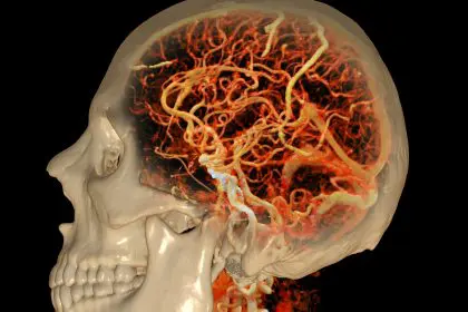
Your eyes might be the window to your future brain health, and what researchers are discovering could change how we detect Alzheimer’s disease forever. Long before memory problems become noticeable, subtle changes in vision and eye function may signal that something significant is happening in the brain.
This groundbreaking connection between eye health and Alzheimer‘s disease represents one of the most promising developments in early detection research. Unlike traditional cognitive tests that can only identify problems after symptoms appear, eye-based assessments could potentially spot warning signs years or even decades before memory loss begins.
The implications are enormous for millions of families who watch loved ones struggle with Alzheimer’s progression. Early detection could mean earlier interventions, better planning, and potentially more effective treatments when they become available.
The retina connection reveals brain secrets
The retina, located at the back of the eye, shares a unique relationship with the brain that makes it an ideal window into neurological health. This thin layer of tissue contains nerve cells that are directly connected to the brain through the optic nerve, making it essentially an extension of the central nervous system.
- Retinal blood vessels mirror brain blood vessels, showing similar changes
- Nerve fiber layers in the retina reflect the health of brain tissue
- Protein deposits in the eye may indicate similar accumulations in the brain
- Blood flow patterns in the retina can reveal circulation problems affecting the brain
What makes the retina particularly valuable for Alzheimer’s detection is its accessibility. Unlike brain tissue, which requires invasive procedures or expensive imaging to examine, the retina can be viewed directly through routine eye exams using relatively simple equipment.
Recent technological advances have made retinal examination even more precise. High-resolution imaging can now detect microscopic changes in retinal structure and function that would have been impossible to see just a few years ago.
Vision changes that raise red flags
The eye changes associated with Alzheimer’s risk go far beyond simple vision problems that might be corrected with glasses. These are subtle alterations in how the visual system processes information and responds to different stimuli.
Color perception difficulties often appear years before other symptoms. People who later develop Alzheimer’s may struggle to distinguish between similar colors, particularly blues and greens. This isn’t about needing stronger reading glasses; it’s about the brain’s changing ability to process visual information.
Contrast sensitivity problems create challenges in distinguishing objects from their backgrounds. Someone might have trouble seeing a white plate on a light-colored tablecloth or difficulty navigating stairs because they can’t clearly see where one step ends and the next begins.
Depth perception changes can make simple tasks like reaching for objects or walking down stairs feel uncertain and dangerous. These spatial processing difficulties reflect changes in brain regions responsible for interpreting three-dimensional information.
Motion detection problems may cause someone to have trouble following moving objects with their eyes or feeling disoriented when things move in their peripheral vision. This can make activities like driving or walking in crowded areas particularly challenging.
Structural changes tell the story
Advanced imaging techniques can now detect physical changes in eye structure that correlate with brain health. These modifications occur at the cellular level and often precede any noticeable symptoms by years.
Retinal nerve fiber layer thinning occurs when the nerve cells that connect the eye to the brain begin to deteriorate. This thinning can be measured with precision and appears to correlate with cognitive decline patterns.
Blood vessel changes in the retina mirror what’s happening in brain blood vessels. Narrowing, irregularities, or changes in blood flow patterns can indicate circulation problems that affect brain function.
Protein accumulations in the eye may reflect similar deposits forming in the brain. These protein clusters, invisible to the naked eye, can be detected through specialized imaging and may indicate the early stages of Alzheimer’s-related brain changes.
Optic nerve changes can signal problems with the connection between the eye and brain. Swelling, color changes, or structural alterations in the optic nerve may indicate neurological problems developing elsewhere.
The sleep and eye connection
Sleep disturbances are common in Alzheimer’s disease, and the eyes play a crucial role in regulating sleep patterns. Changes in how the eyes respond to light and darkness can disrupt the body’s natural circadian rhythms, leading to sleep problems that may appear years before memory issues.
The pupils’ response to light changes can reveal problems with the autonomic nervous system, which controls many automatic body functions. Slower or irregular pupil responses may indicate neurological changes affecting brain function.
Eye movement during sleep, particularly REM sleep, can be altered in people who later develop Alzheimer’s. These changes in sleep architecture may be detectable through specialized testing long before cognitive symptoms appear.
Light sensitivity changes can affect how well someone sleeps and how alert they feel during the day. Increased sensitivity to bright lights or difficulty adjusting to changing light conditions may signal developing neurological problems.
Reading and visual processing challenges
Long before memory problems become obvious, people at risk for Alzheimer’s may experience subtle changes in how they process written information. These aren’t simple vision problems that can be fixed with stronger glasses, but rather difficulties with how the brain interprets what the eyes see.
Reading speed may slow down gradually, with people taking longer to process the same amount of text they could previously read quickly. This change often goes unnoticed initially because it develops slowly over time.
Tracking problems make it difficult to follow lines of text smoothly. Someone might lose their place while reading, skip lines, or have trouble moving their eyes systematically across a page.
Visual scanning difficulties can make it hard to find specific information in complex visual displays. This might show up as trouble reading maps, following recipes, or finding items in cluttered spaces.
Word recognition problems may cause familiar words to look strange or unfamiliar, even when vision is otherwise clear. This represents a breakdown in the connection between visual input and language processing in the brain.
The technology revolution in detection
Modern eye examination equipment has transformed what doctors can see and measure during routine eye exams. These technological advances make it possible to detect incredibly subtle changes that would have been invisible just a few years ago.
Optical coherence tomography creates detailed cross-sectional images of retinal layers, allowing doctors to measure thickness changes down to microscopic levels. This technology can detect nerve fiber loss long before it would be visible through traditional examination methods.
Retinal photography with specialized filters can highlight protein deposits and blood vessel changes that indicate neurological problems. These images can be compared over time to track progression and identify patterns associated with Alzheimer’s risk.
Eye movement tracking technology can detect subtle changes in how the eyes move and focus. These measurements can reveal problems with attention, processing speed, and executive function that may precede obvious cognitive decline.
Pupil response testing uses controlled light stimuli to measure how quickly and consistently the pupils respond. Abnormal responses may indicate problems with the autonomic nervous system or brainstem function.
Lifestyle factors that affect eye-brain health
The same lifestyle factors that promote brain health also support eye health, creating opportunities for people to potentially reduce their risk of developing Alzheimer’s disease through healthy choices.
Cardiovascular health plays a crucial role in maintaining good blood flow to both the brain and eyes. Regular exercise, healthy diet, and management of blood pressure and cholesterol can help preserve the delicate blood vessels that supply these critical organs.
Sun protection for the eyes may help prevent damage that could accelerate aging-related changes. Wearing sunglasses and wide-brimmed hats can reduce exposure to harmful ultraviolet radiation that may contribute to retinal damage.
Nutrition specifically supports eye and brain health through antioxidants, omega-3 fatty acids, and other nutrients that protect against inflammation and oxidative stress. Foods rich in these compounds may help maintain healthy vision and cognitive function.
Sleep quality affects both eye health and brain function. Poor sleep can accelerate aging-related changes in both systems, while good sleep hygiene supports the natural repair processes that keep these organs functioning optimally.
The future of early detection
As research continues to unveil the connections between eye health and Alzheimer’s disease, the potential for early detection grows more promising. Future eye exams may routinely include screenings for neurological risk factors, making early identification of Alzheimer’s risk as common as checking for glaucoma or diabetes.
Artificial intelligence is being developed to analyze retinal images and identify patterns associated with Alzheimer’s risk. These computer systems could potentially detect subtle changes that human observers might miss, improving the accuracy and consistency of early detection efforts.
Home monitoring technology may eventually allow people to track their own eye health and receive alerts about changes that warrant professional evaluation. This could make regular monitoring more convenient and accessible for people at risk.
Integration with other health data could create comprehensive risk profiles that combine eye health information with genetic testing, cognitive assessments, and other biomarkers. This holistic approach could provide more accurate predictions about individual Alzheimer’s risk.
The eyes truly may be the windows to the soul, and in the case of Alzheimer’s disease, they’re becoming windows to the future of brain health. As our understanding of these connections continues to grow, the simple act of getting an eye exam may become one of the most important steps people can take to protect their cognitive future.


